Fichier:Anaplastic astrocytoma.jpg
Gréisst vun dëser Duerstellung: 800 × 552 Pixel. Aner Opléisungen: 320 × 221 Pixel | 640 × 442 Pixel | 1.024 × 707 Pixel | 1.200 × 828 Pixel.
Original-Fichier (1.200 × 828 Pixel, Fichiersgréisst: 200 KB, MIME-Typ: image/jpeg)
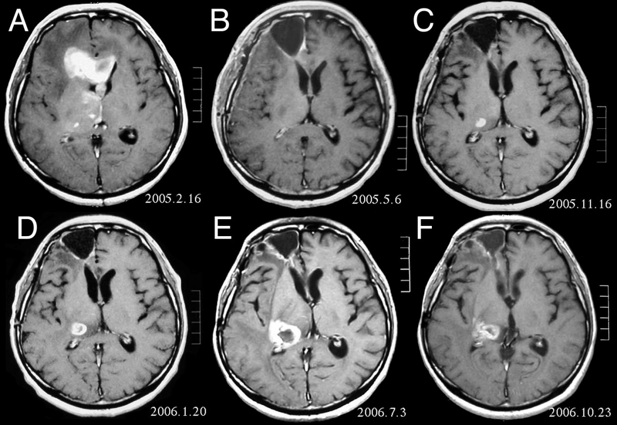
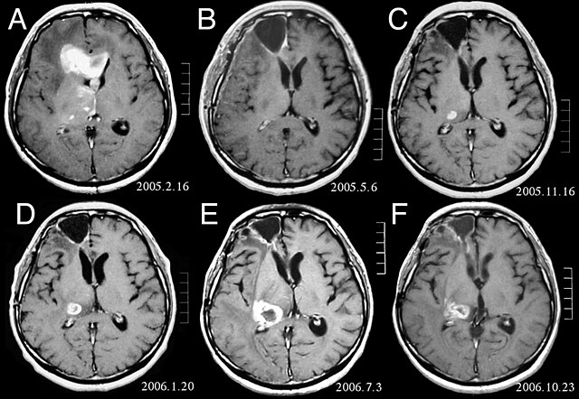
| Beschreiwung | MRI of brain. (A) Initial MRI on February 16, 2005, shows a tumor in the right and left frontal lobe as well as the right thalamus. (B) MRI after surgery, radiation and chemotherapy. The tumor has completely disappeared except for slight enhancement adjacent to the surgical margin. (C) Recurrence of the thalamic tumor despite maintenance chemotherapy on November 16, 2005. (D) Increase in size of the thalamic tumor two months after stereotactic radiotherapy. (E) After 6 cycles of TMZ therapy, the thalamic lesion enlarged, and the patient developed dysarthria and hemiparesis. (F) After 2 courses of treatment with interferon-beta and TMZ, the tumor shows a partial response. |
| Datum | |
| Quell | Fujimaki T, Ishii H, Matsuno A, Arai H, Nakagomi T.Effectiveness of interferon-beta and temozolomide combination therapy against temozolomide-refractory recurrent anaplastic astrocytoma.World J Surg Oncol. 2007 Aug 4;5:89. PMID 17683572 doi:10.1186/1477-7819-5-89 |
| Auteur | Fujimaki T, Ishii H, Matsuno A, Arai H, Nakagomi T. |
| Autorisatioun (Dëse Fichier nach eng Kéier benotzen) |
BioMedCentral License |
Dëse Fichier ass ënner der Creative Commons Attribution 2.0 Generic Lizenz disponibel.
- Dir kënnt:
- D'Wierk deelen – kopéieren, verdeelen a weiderginn
- D'Wierk kombinéieren – adaptéieren
- Ënner dëse Konditiounen:
- Attributioun – Dir musst appropriéiert Informatiounen iwwer den Auteur uginn, e Link op d'Lizenz maachen, an uginn ob Ännerunge gemaach goufen. Dës Informatioune kënnen op eng räsonabel Manéier gi ginn, awer ouni datt den Androck entsteet datt deen deen d'Lizenz ginn huet Iech oder Är Benotzung approuvéiert oder ënnerstëtzt.
Versiounen
Klickt op e bestëmmten Zäitpunkt fir déi respektiv Versioun vum Fichier ze kucken.
| Versioun vum | Miniaturbild | Dimensiounen | Benotzer | Bemierkung | |
|---|---|---|---|---|---|
| aktuell | 16:47, 25. Feb. 2008 | | 1.200 × 828 (200 KB) | Filip em | {{Information |Description=MRI of brain. (A) Initial MRI on February 16, 2005, shows a tumor in the right and left frontal lobe as well as the right thalamus. (B) MRI after surgery, radiation and chemotherapy. The tumor has completely disappeared except f |
Benotze vu Fichieren
Dës Säit benotzt dëse Fichier:
Globaalt Benotze vum Fichier
Dës aner Wikie benotzen dëse Fichier:
- Benotzt op ar.wikipedia.org
- Benotzt op bg.wikipedia.org
- Benotzt op cs.wikipedia.org
- Benotzt op da.wikipedia.org
- Benotzt op de.wikipedia.org
- Benotzt op el.wikipedia.org
- Benotzt op en.wikipedia.org
- Benotzt op es.wikipedia.org
- Benotzt op et.wikipedia.org
- Benotzt op hi.wikipedia.org
- Benotzt op hr.wikipedia.org
- Benotzt op hu.wikipedia.org
- Benotzt op it.wikipedia.org
- Benotzt op kk.wikipedia.org
- Benotzt op lv.wikipedia.org
- Benotzt op mk.wikipedia.org
- Benotzt op mt.wikipedia.org
- Benotzt op nl.wikipedia.org
- Benotzt op no.wikipedia.org
- Benotzt op pl.wikipedia.org
- Benotzt op ro.wikipedia.org
- Benotzt op sk.wikipedia.org
- Benotzt op sl.wikipedia.org
- Benotzt op sq.wikipedia.org
- Benotzt op sr.wikipedia.org
- Benotzt op uk.wikipedia.org